Most bioseparation and detection tools used in the laboratory employ slab gel-based electrophoresis technologies, which have routinely been used for the analysis of biomolecules (i.e., DNA, RNA, protein, carbohydrates, etc.) since their inception more than 30 years ago. However, slab gel electrophoresis for bioanalysis is a labor-intensive technique. Further, the method needs to be drastically improved in terms of throughput, resolving power, and cost per sample run.
A microfluidic approach to gel electrophoresis (microchannel device to simplify gel electrophoresis), capillary gel electrophoresis (CGE) offers numerous applications.1‒5 Benefits of CGE technology are its high resolution, high sensitivity, and reliability.2,3 Although it enjoys wide acceptance in the biotech industry, CGE is sometimes avoided in routine analysis due to its reputation for being high in cost and having high failure rates. However, this is no longer the case—the Qsep1 portable CGE instrument from BiOptic (New Taipei City, Taiwan [R.O.C.]) is cost-effective and operates in wireless mode using WiFi.6‒8 The present iteration of the instrument provides simplified, high-efficiency, highly sensitive results; is cost-effective; and uses WiFi-type communication for biomolecule detection applications. The system is a miniaturized version of the Qsep100 system vertical capillary gel electrophoresis system, which can complete 4‒16 samples analysis.
The portable CGE-based bioseparation system (Figure 1) measures 15 × 15 × 20 cm and weighs 5 kg. The system is configured to conduct bioseparations in a glass capillary separation channel (microfluidic) using a Smartphone app to operate via WiFi.
 Figures 1 ‒ a) and b) The Portable CGE Instrument (Qsep1) with two-axis sample tray. The pen-shaped gel- cartridge beside the instrument shows the relative size.
CGE detection
Detection for the portable CGE system is light emitting diode-induced fluorescence (LED-IF) using optical fibers for both excitation and emission detection. Fluorescence is a spectrophotometric method of analysis in which the molecules of the analytes are excited by irradiation at a certain wavelength and emit radiation at a different wavelength. The emission spectrum provides information for both qualitative and quantitative analysis. Generally, the advantage of fluorescence detection over absorbance detection is the superior detectability (detection sensitivity). For efficient fluorophores, single-molecule detection in small volumes has been demonstrated. 2,3 This is in part because the fluorescence signal is measured against a relatively dark background as a result of the emitted radiation being detected at a wavelength that is different from the wavelength of the incident radiation (e.g., the wavelength of the emitted fluorescence is at longer wavelengths than the excitation radiation).
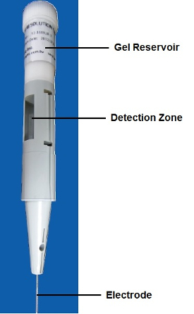 Figure 2 ‒ Pen-shaped gel cartridge.
In the portable CGE system, the fluorescence emission signals produced by the separated analytes are collected at the detection zone of the capillary using an optical fiber, which is then relayed to the detector module (using PM, Si, SiPMT, or CCD-type detectors) with a built-in emission filter (band-pass or long-pass filter) for FITC, EtBr, or other related fluorescence detection applications.
CGE system
The system is composed of a fully automated, two-axis sample tray mechanism that interfaces with the pen-shaped gel-cartridge (Figure 2). The gel cartridge is inserted from the top inside the instrument and detection optical fibers are latched manually by two control knobs to complete the optical connections (excitation and emission) to the detection zone of the gel cartridge. The sample tray can accept standard PCR wells (4‒8 wells), and is designed to accept and analyze 4‒16 samples from standard PCR wells or BiOptic designed 8-Micro-Wells. An additional four sample wells are used for the park, dip, and wash steps for capillary tip cleaning and a buffer reservoir for high-voltage separation purposes.
System operation and results
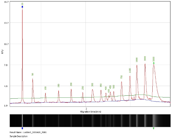 Figure 3 ‒ Separation of DNA ladder including the two alignment markers (lower marker = 20 bp; upper marker = 5000 bp). Both an electropherogram and a gel image are shown.
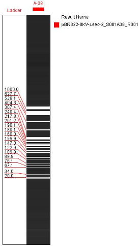 Figure 4 ‒ Gel image. Separation of DNA ladder (8-kV separation with 20- and 1000-bp alignment markers).
The portable CGE system accepts a single-channel, pen-shaped gel cartridge (Figure 2) that has an integrated top gel/buffer reservoir. The instrument is directly coupled to an external air-pressure source (i.e., portable pressure air pump, syringe pump, or N2 gas tank) that provides air or N2 pressure to fill up the capillary (microfluidic channel) with the separation gel/buffer from the top gel reservoir of the cartridge. Depending on the viscosity of the separation gel/buffer, pressures of up to 40‒60 psi can be manually applied to the capillary through the top buffer reservoir by the air-pressure source.
The cartridge reservoir is equipped with built-in electrode (anode) that is automatically connected to the highvoltage power supply for electrophoresis when installed inside the instrument. The test samples are introduced to the separation capillary (microfluidic channel) by electrokinetic injections from the cathode end of the gel cartridge using the control panel (Android operating devices such as a Smartphone app). A high-voltage power supply is used to deliver 0‒8 kV of electrical field to the capillary for the electrokinetic injection and separations (typically 4‒8 kV) of biomolecules. The negatively charged samples (i.e., DNA) are injected by applied high voltage (1‒4 kV) into the gel cartridge, and vertical electrophoresis of the biomolecule is achieved by the portable CGE.
The system is capable of high-resolution detection of PCR products in 60‒300 sec in the range of 20‒5000 bp with a resolution of 2‒4 bp below 500 bp (Figures 3‒5).
 Figure 5 ‒ High-resolution detection of PCR products at 269‒272 bp with the lower and upper markers.
Smartphone operation/WiFi communication
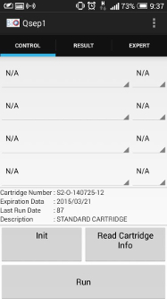 Figure 6 ‒ Smartphone app main control.
BiOptic has developed a Smartphone app that has the ability to operate the Qsep1 remotely via WiFi (Figure 6).
Launching the Smartphone app for system operation
- There are several steps to establish communications via Smartphone and start operating the Qsep1 instrument:
- Turn instrument on
- Launch phone app
- Scan QR code (bar code) in the front of the instrument using the Smartphone
- Automatic handshake (communications established)
- Start system operation:
a. Install and latch gel cartridge inside the instrument
b. Position the sample well inside the sample tray
c. Launch method
d. Select sample tray position
e. Run separation and data collection
f. Display results (gel image at the app and cloud computing with the SD card)
g. Analyze data at remote computer by transferring the data from the SD card physically or via the cloud (see Figure 7).
Conclusion
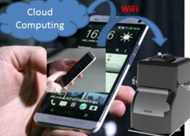 Figure 7 ‒ Wireless operation of Qsep1 via Smartphone with cloud computing feature.
A very simple and portable CGE instrument provides rapid and cost-effective bioseparation and detection for research and clinical use. The simplified CGE instrument design produces accurate, precise, and robust results for post-PCR (DNA fragment analysis) or genotyping applications. The fully automated portable CGE system is operated remotely (wireless) with Android operating devices (i.e., Smartphones and tablets, etc.) using the BiOptic Smartphone app to complete a 4‒16 sample run. The completed electropherogram or gel image results are then displayed on the Smartphone app or can be retrieved via an SD card to be transferred to a computer or via cloud computing for further data analysis (postprocessing) using BiOptic Q-Analyzer software.
References
- Guttman, A.; Cook, N. Capillary gel affinity electrophoresis of DNA fragments. Anal. Chem. 1991, 63, 2038‒42.
- Amirkhanian, V.D.; Liu, M.-S. Low-cost and high-throughput multi-channel capillary electrophoresis (MCCE) system for DNA analysis. Biomedical nanotechnology architectures and applications. Proceedings of SPIE 2002, 4626, 238–45.
- Liu, M.-S.; Amirkhanian, V.D. DNA fragment analysis by an affordable multiple-channel capillary electrophoresis system. Electrophoresis 2003, 24, 93‒5.
- Huang, X.C.; Quesada, M.A. et al. DNA sequencing using capillary array electrophoresis. Anal. Chem.1992, 64, 2149–54.
- Clark, S.M.; Mathies, R.A. High-speed parallel separation of DNA restriction fragments using capillary array electrophoresis. Anal. Biochem. 1993, 215, 163–70.
- Shimadzu News. Expanding HPLC Network Functionality: Improved Efficiency with New iPad Control, 10/1/2014; http://www.shimadzu.com/about/pressrelease/5iqj1d0000011yva.html
- Shimadzu News. New LabSolutions Direct Function Enables HPLC Access from Smartphone or Tablet PC—provides a remote operating environment for operations such as HPLC control, starting analysis and analysis monitoring, 8/29/2013; http://www.shimadzu.com/about/pressrelease/5iqj1d000001v9lw.html
- Li, B.; Li, L. et al. A smartphone controlled handheld microfluidic liquid handling system. Lab-on-a-Chip 2014, 14, 4085‒92.
Eric Tsai, Allen Chi, Myron Yu, and Hsinhan Huang are with BiOptic, Inc., New Taipei City, Taiwan (R.O.C.). Varoujan Amirkhanian is with BiOptic, Inc., 1409 1/2 Foothill Blvd., La Canada Flintridge, CA 91011, U.S.A.; tel.: 818-679-4413; e-mail: [email protected]; www.bioptic.com.tw