Despite a blizzard in the Eastern United States, 6293 scientists—an increase of about 25% from the conference’s previous high—were able to travel to sunny San Diego to discuss laboratory automation and screening at SLAS2016, held January 24–27. The intensity of interest in automation and biological assays, reflected in both attendance and the content of 140 lectures and 400 posters, validates the symbiotic combination that drove the merger of the Society for Laboratory Automation and the Society for Biological Screening in 2010.
Cell biology
The keynote lecture by Dr. Michael Gottesman of the Laboratory of Cell Biology, National Cancer Institute (Bethesda, Md.), set the stage for the meeting. Focusing on drug resistance in cancer, he discussed the experimental difficulty in dealing with cell growth in a dynamic solid tumor, and as an example cited oxygen tension. When a cell becomes cancerous it can rob nutrients, such as oxygen, from neighboring cells. As they grow, high metabolism cancer cells proliferate and interior cells suffer from oxygen starvation since the exterior cells use it first. Waste products build up in the interior and may poison the central cells, unless these cells develop countermeasures, such as alternate energy pathways.
Such variations in cell environment, as well as variations caused by metabolic processes, may explain why angiogenesis as a cancer treatment has not worked as well as initially hoped. Gottesman noted that typical in vitro cell biology experiments involve 21% oxygen concentration. Yet, in vivo, oxygen tension varies significantly. This is one example of why there is difficulty translating in vitro results to in vivo systems.
Dr. Robin Felder of the University of Virginia (Charlottesville) provided a list of needs for cell therapy: large-scale expansion of cells; ability to obtain data that is relevant and useful in triage and therapy of humans; and culturing cells in 3-D, which appears to be much more useful than 2-D for providing clinically relevant data. Professor Felder also saw the need for fully automated cell culture systems that include programmed temperature and oxygen tension to control cell growth. From Dr. Felder’s remarks, it appears that laboratory-scale cell biology needs to improve before cell therapy meets the efficacy and safety expectations of the public.
Cell preservation
I came away from the conference impressed with the improvements in protocols and apparatus for cell biology, which I see as being partly driven by impending advances in cell therapeutics and by advances in bio-banking and supply-chain management. For example, in the Brooks Automation (Chelmsford, Mass.) booth, a major part of the backdrop focused on controlling temperature in the steps from bio-banked specimen cell therapeutic delivery to a patient. Successful freezing and thawing of cells minimizes formation of ice crystals, which can damage cell structure, reducing viability. For example, GLP is to thaw samples by bringing them to a temperature of 37 °C in 60–90 seconds. Common practice has been to float the vial in an ice bath, which thaws the cell sample slowly. But slow thawing gives water time to enter the cold, dehydrated cells and form ice crystals. The crystals damage cell membranes and organelles, resulting in cell death.
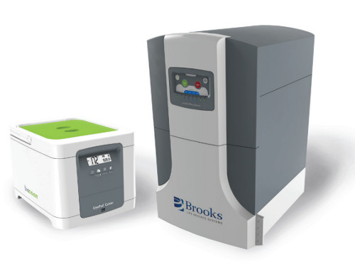 Figure 1 – CryoPod carrier from Brooks Automation.
The Brooks CryoPod carrier (Figure 1) allows sample or product transport at <–150 °C. For use in transporting frozen cells to the lab bench or bedside, it holds samples below the Tg (glass transition temperature) of water (–135 °C) for 20 days. Brooks also cooperates with BioCision (San Rafael, Calif.), a company that provides a programmable melting module that heats sample tubes to 37 °C in 60–90 seconds without overshoot. All of this minimizes the variable of thermal stress imposed upon samples during bio-banking.
Most biological samples are preserved by freezing. One can avoid repeated freeze thaw cycles by aliquotting a master prep into small vials at volumes suitable for the intended assays. But this increases the storage volume in the freezer. Glenn Smith of CryoXtract Instruments (Woburn, Mass.) proposed aliquotting frozen specimens directly with a micro-coring device that operates directly on the frozen samples, including tissue and frozen liquids. Using Automated Frozen Aliquotting Technology, scientists can drill a small core out of a frozen specimen in a vial. The frozen aliquot is delivered to the sample tube, where it can be quickly heated, extracted, filtered and diluted to volume. Typical samples include urine, blood and feces. Screening cell lines for best conditions for transfecting CRISPR plasmid libraries was the topic of a poster by Drs. Anna Lina Cavallo and Silvio Di Castro of AstraZeneca (Gothenburg, Sweden). Loading plates took too long, which reduced cell viability. With manual preparation, cell viability for the first wells differed from similar controls in the last wells. Automating plate filling with an Echo 555 liquid handler (LabCyte, Sunnyvale, Calif.) with OMICS S2 calibration solved the problem. Some reagents required dilution for effective dispensing with the Echo. However, once optimized, it was possible to quickly select the most effective transfection reagents and optimize the concentration.
Factors affecting data quality
Interest in data quality was a commonly expressed concern and, of course, automation—reducing human involvement— provided the biggest improvements. The density of a 384-/1536-well plate taxes the limits of human capability. Yes, humans can load every well in the plate with a common reagent volume, but loading different volumes into specific wells becomes problematic. Uncertainty increases with the number of wells.
Automated handheld pipetting
 Figure 2 – VIAFLO ASSIST from INTEGRA Biosciences.
The VIAFLO ASSIST (Figure 2) from INTEGRA Biosciences (Hudson, N.H.) automates pipetting into multiple-well plates. Electronic pipets can be programmed so that they can be held vertically, prewetting the tip by two or three aspirate-and-dispense cycles. Tip immersion depth can be controlled to improve precision and reduce carryover. Mixing cycles are programmable in height, velocity and cycle number. ASSIST does not skips rows or wells, and users attest that it provides an economical means to improve assay precision.
Digital dispensing of 11-pL to 10-μL volumes
For several years, scientists have wondered if direct digital dispensing technology used in inkjet printers would be adaptable for liquid dispensing. Hewlett-Packard (Corvallis, Ore.) provided a short answer: Yes! In an apparent OEM arrangement, Tecan (Mannedorf, Switzerland) rebranded the HP D300e to the Tecan D300 E, with which a dose from 11 pL to 10 μL can be dispensed in any well. This facilitates direct measurement of dose response curves without serial dilution, which is vulnerable to compounding errors. Loading a plate is generally faster, too: all that’s needed is to insert a disposable dispense head and load a small reservoir and it’s ready to run. Working with such small volumes, expensive reagents are dispersed over many more wells, reducing costs.
High-throughput screening gets faster and faster
Over the last 10 years, high-throughput screening (HTS) has been conducted at ever-increasing speeds, reducing per-well dispense time from a few minutes to a few seconds to a well per second, as reported by Dr. Melanie Leveridge of GlaxoSmithKline (Brentford, U.K.). The biggest factor in HTS time improvements came from replacing chromatography with higher-speed matrix-assisted laser desorption ionization-time of flight mass spectrometry (MALDI-TOF MS) with multiple reaction monitoring. These instruments have the specificity needed to identify fragment peaks. Smaller wells, which increase well-to-plate ratios, contribute to the method’s speed. The small wells require handling nanoliter volumes of liquids. Dr. Leveridge reported use of 1536-well plates and some runs with 6144-well plates, while other lecturers select 384 wells as more practical, given the abilities of contemporary liquid-handling modules.
Replacing the LC step with laser desorption is also economically favorable. According to one author, replacing a five-minute UHPLC run with MALDI-TOF reduced assay costs from about $540/sample to less than $1.00/sample. Other presenters corroborated crossing the dollar/well threshold with MALDI-TOF.
A poster by E. Hall of LabCyte and colleagues at AstraZeneca (Macclesfield, U.K.) and IonSense, Inc. (Saugus, Mass.) described one system. In the example, a modified Echo liquid handler ejects 2.5-nL drops of sample from a 384-well plate onto IonSense’s Vapur gold substrate mesh. Excited helium from the DART (Direct Analysis in Real Time)-MS interface from IonSense impinges on the gold mesh, which vaporizes and ionizes the sample. Throughput is three samples per second. The interface was evaluated using an assay of caffeine in water in a 384-well plate. CV for the plates was less than 15% using single-ion monitoring. The authors went on to use the system in a 1000-compound screen.
The move to 1536 and 6144 wells
There seemed to be a clear acceptance that 1536-well plates held a place in the mainstream. A lecture by Dr. Helen Plant of AstraZeneca (London) reported her experience in moving from 384 to 1536 wells/plate for phenotypic assays. Until recently, limitations in liquid handling, including washing and media exchange, precluded the use of 1536-well plates. But she was attracted to the higher-density plates due to lower reagent cost and expected higher throughput.
The washing issue was addressed by the BlueWasher from Blue Cat Bio (Toronto, Canada). The device uses centrifugation for noncontact, contamination-free removal of liquids from all plate formats, including 1536-well, and achieves near-zero residual volume in each well after washing. Users can add up to four separate wash solutions, which are often required for some complex phenotypic assays.
Dr. Plant’s bridging study successfully compared EC50 data generated in both 384- and 1536-well formats. Then, for further confirmation, drug master screens of a 43K library were run in 1536-well plates, which required only two weeks. Required number of cells was reduced 25%, reagent cost/ assay was now deemed affordable for screenings at scale, and there was a fourfold reduction in the campaign time. During post-lecture Q&A, Plant was asked if she expected to eventually use 6144-well plates. She replied that evaporation is so rapid at 1536 that higher open-well densities may not be practical and microfluidic tubes will replace wells.
Trueness replaces accuracy
Dr. George Rodrigues of Artel (Westbrook, Maine) discussed recent updates to international standards for liquid handling. The most significant change from his working group recognized that the term “accuracy” is ambiguous, and so “trueness” was chosen as a substitute. Precision will still measure scatter of assay results, and “trueness” will measure the difference between the measured value and the true value.
Notable new products
SLAS organizers recognize that innovative new products often originate in small firms that cannot afford or even staff a regular booth. These firms (such as SiO2 Medical Products and Click Bio, below) were given an opportunity to engage the scientific community through the AveNEW section on the exhibition floor. It consisted of a 20 × 20-sq.-ft. booth in the middle of the hall.
SiO2 Medical Products (Auburn, Ala.) exhibited plasticware incorporating an inert silica layer that is bonded using plasma-enhanced chemical vapor deposition to items such as polyolefin or polystyrene plastic vials or multiple-well plates to improve storage stability, as required in source plates for compound libraries. Vapor of hexamethyldisiloxane is swept into the plastic well and destroyed with oxygen plasma. A thin layer of silica is deposited onto the plastic surface. Surprisingly, the silica is very flexible and resistant to cracking or spalling. The silica film stops the migration of oxygen and water through the plastic. If a hydrophobic surface is desired, the silica can be reacted with silanes, which makes a polar surface nonpolar.
More than half of the instruments in the exhibition were designed to process plates manufactured to ANSI SLAS 1-2004 standards. Click Bio (Reno, Nev.) introduced the Click plate, which facilitates designing and assembling various reservoirs and vials that fit into the 1–2004 format. The components fit together and then click into the ANSI-standard plate perimeter. This design can be used to avoid edge effects in cell culture, or to isolate reagents and waste.
Consistent measurement of cell confluence is essential to cell culture QC and signal normalization. Tecan introduced the Spark20M microplate reader for label-free cell-analysis imaging for cell counting and viability analysis, especially during long run assays. Spark software enables the scientist to count specific locations or regions in the well with either kinetic or endpoint modes. Since it is label-free, complications from labels and transfection are avoided.
Second harmonic generation
Small-molecule drugs can change the higher order structure of target proteins. Biodesy, Inc. (South San Francisco, Calif.) introduced a second harmonic generation (SHG) platform that measures ligand-induced conformational changes. Biodesy explained that SGH arises when a protein receptor is covalently labeled with a fl uorescent dye via an amine or thiol linker. The protein is tethered to a lipid bilayer on a detection cell via a histamine tag. The dyes generate second harmonic light when illuminated with a femtosecond laser. Fluorescence intensity is a strong function of the average angular orientation of the dye. Even sub-Angstrom shifts produce a detectable signal change. SHG is insensitive to mass or primary structure. Biodesy reported that SHG is suitable for testing potential ligands, including antibody/antigen interactions.
Dr. Charles Wartchow of the Novartis Institutes for Biomedical Research (Cambridge, Mass.) evaluated the SHG platform for binding peptide and protein fragments. Wartchow pointed out that the SHG is different from more traditional binding assays such as surface plasmon resonance (SPR), since the binding must involve a change in the conformation of the target. Wartchow and colleagues were interested in evaluating drug libraries, and thought that the SHG-sensitive conformational change could be related to a possible mechanism of action (MOA). In a blinded study, results obtained for SHG were compared with 2-D nuclear magnetic resonance (NMR) and X-ray crystallography. A Venn diagram of three overlapping circles (one each for SHG, NMR and X-ray) had significant numbers in all regions, which suggests that each technique is complementary and useful in lead discovery. Wartchow concluded that SHG is useful in screening fragments with therapeutic targets that induce conformational change.
SLAS2017 will be held February 4–8 at the Walter E. Washington Convention Center in Washington, D.C. Monitor www.SLAS2017.org for the latest information.
Robert L. Stevenson, Ph.D., is Editor Emeritus, American Laboratory/Labcompare; e-mail: [email protected]