An articulated opto-mechanical arm, the daVinci arm (Harrick Scientific Products, Pleasantville, NY) (Figure 1), was developed to enable in situ analysis of large samples.1 It permits the analyses of samples by specular reflection, diffuse reflection, and internal attenuated total reflecion (ATR) techniques. It mounts into the sample compartments of most commercial FTIR spectrometers. The ability to use a deuterated triglycine sulfate (DTGS) detector allows spectra to be recorded below 650 cm–1. Since the arm is articulated, samples can be analyzed within a range of distances in front, below, or above the sample compartment at a wide range of angles. The integral camera provides for magnified viewing and image capture of the sampled spot. The entire optical path of the IR beam is enclosed and integrated into the purge of the host spectrometer.
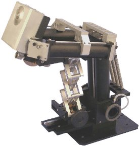
Figure 1 - daVinci arm.
In specular and diffuse reflection mode, the sampled spot is about 1.25 mm in diameter. In ATR mode, the spot is 0.5 mm in diameter. The live image provided by the integral camera serves as a guide for placing the sample at the focus of the IR beam.
In this work, the daVinci arm was used for the analysis of a large (26 in. high × 22 in. wide) oil canvas. The painting was too large to fit into the sample compartment of a spectrometer and could not be cut or otherwise damaged by the analysis. The authors demonstrate that the daVinci arm enables the acquisition of good-quality spectra outside of the sample compartment. Since the artwork itself could not be touched, the only spectroscopic technique that could be utilized was diffuse reflection, which is a noncontact method.
Optically, the daVinci arm works as follows. The infrared beam is directed by the optics in the sample compartment of the spectrometer through the arm to the sampling spot some distance away from the sample compartment. There it interacts with the sample either by a noncontact reflection technique or by diamond ATR, a contact sampling method. After interaction of the incident beam with the sample, the light reflected from the sample is returned to the sample compartment and directed to the spectrometer’s detector.
Since the light transfer is accomplished entirely by front-surface aluminum mirrors within purge-containing tubes, there is only a moderate loss of light intensity due to the transfer optics. Unlike fiber optics, which can also be used for sample analysis outside of the sample compartment, there are no spectral range limitations. The arm can be used from the UV to the far-IR.
Any flat or convex-shaped object can be placed in front of the spectrometer for analysis. For sheets of paper, textiles, and similar materials, placing them flat on a table and bringing the arm in from the top is the easiest configuration for analysis. For other materials, such as the painting studied in this work, placing the sample vertically in front of the spectrometer is the most convenient position. In all cases, it is necessary to have the means to manipulate the sample so that the desired spot on the sample can be analyzed.
The arm is designed such that, once aligned, it stays aligned within the entire available range of motion. In practice, some reoptimization of the optical alignment may be needed when the position of the arm is changed. It is best to collect the background spectrum in a position that is close to the final sampling position.
When used in the ATR mode, the arm is totally enclosed, and the purge gas within the arm is fully contained. However, when used in diffuse/specular mode, the light must exit the arm, interact with the sample, and return back. An optically transparent window could be used to enclose the purged environment of the arm, but that would limit the spectral range, introduce reflectance losses, and defocus the beam. To avoid using an optical window, the arm uses a purge shroud that leaves an aperture for light to exit and return. The size of this opening is as small as possible, making it feasible to maintain a positive purge pressure inside the arm by slightly elevating the pressure of the purge gas.
Experimental
All spectra were collected at 8 cm–1 resolution with 64 scans coadded. For the collection of the background spectrum, a piece of machined stainless steel was used as a reference. The background spectrum was collected at the beginning of the experiment. For the last measurement, the position of the sampling head of the arm moved significantly; thus the alignment was reoptimized by maximizing the signal on the detector.
Results and discussion

Figure 2 - Artwork shown in the easel used for positioning.

Figure 3 - Close-up with analyzed points indicated.
The painting analyzed is shown in Figure 2. The painting was placed on an easel that allowed the painting to be tilted, raised, and lowered albeit in coarse steps of about 4 in. The tilt of the easel was adjusted to make the canvas vertical and eight points were selected for analysis, as indicated in Figure 3. The choice of points required moving the painting sideways as well as up and down. Since the easel enabled only coarse vertical adjustments, the daVinci arm was moved vertically to reach the chosen points. Each time the canvas was moved, the sampling head of the arm was retracted to avoid inadvertently damaging the artwork. After the canvas was brought into the desired position, the sampling head of the arm was extended toward the canvas into its sampling position.

Figure 4 - daVinci arm shown in position analyzing point H.
The positioning of the arm relative to the sample had to be done very carefully, since the shroud had to be brought very close (1 mm) to the sample without actually touching it. Figure 4 shows the arm in position (point H) acquiring the spectrum shown in Figure 5. Significant differences are seen in the diffuse reflectance spectra measured from different locations on the painting, as shown in Figures 5–12. Common features are presumably due to the common oil matrix and perhaps the underlying canvas, while differences are due to the various colored pigments used. However, these spectra are not straightforward to interpret. An oil painting is a complex reflecting system. For a detailed analysis, a careful study could be conducted to analyze and identify spectral features that are due to oil matrix, canvas, varnish, different pigments, etc. Once these samples analyzed by diffuse reflectance are thoroughly characterized, and the effects of the various components identified, the spectra could be more readily interpreted.

Figure 5 - Diffuse reflectance spectrum of point H with a photograph of the area examined.

Figure 6 - Diffuse reflectance spectrum of point A.

Figure 7 - Diffuse reflectance spectrum of point B.

Figure 8 - Diffuse reflectance spectrum of point C.

Figure 9 - Diffuse reflectance spectrum of point D.

Figure 10 - Diffuse reflectance spectrum of point G.

Figure 11 - Diffuse reflectance spectrum of point F.

Figure 12 - Diffuse reflectance spectrum of point E.
Note that although the spectra were recorded outside of the spectrometer sample compartment, the spectral quality is comparable to that of typical incompartment measurements.
Conclusion
It has been demonstrated that, using the daVinci arm, good-quality spectra can be collected from spots on a large, flat object that otherwise could not be analyzed by FTIR spectroscopy. The particular object analyzed in this work was an oil painting that could not be touched while analyzed. Thus, diffuse reflection, a noncontact technique, was used to conduct the analysis. Presenting the object for analysis required moderate effort and care but was not difficult or time consuming. Less than 5 min was needed to get the artwork positioned for each measurement. Virtually any point on the sample could be analyzed.
The diffuse reflectance spectra obtained show considerable detail and thus contain significant information. This information is not easily interpreted, but the spectra do contain valuable information about the sample that could not be obtained otherwise.
Reference
- Milosevic, M.; Milosevic, V.; Padron, L.; Wang, J.; McGlinchey, C. IRUG 7, MOMA, New York, 2006.
Dr. Berets, Mr. Lucania, and Mr. Christenson are with Harrick Scientific Products, 141 Tompkins Ave., 2nd Floor, Box 277, Pleasantville, NY 10570, U.S.A.; tel.: 914-747-7209; fax: 914-747-7209; e-mail: [email protected]. Mr. M. Milosevic and Mrs. V. Milosevic are with MeV Photonics, Westport, CT, U.S.A.