The city of Philadelphia welcomed the American Society for Cell Biology (ASCB) for its 50th Annual Meeting, held from December 11 to 15 (Figure 1). The meeting featured the new tagline, The Science of Life, the Life of Science, underscoring the Society’s commitment to advancing discovery in the field of cell biology research. Philadelphia was a fitting host city—It boasts several key science attractions of its own, including the Academy of Natural Sciences, one of the oldest natural science research institutions, and the Franklin Institute, one of the oldest centers for science education and development.
Figure 1 - The Pennsylvania Convention Center (Philadelphia, PA). Photo by Jim McWilliams for the PCVB.
Key topics
The ASCB Annual Meeting highlighted emerging research interests and fast-breaking technologies, including super-resolution microscopy and mathematical modeling. Some topics of interest at the meeting included membrane and organelle biology, focusing on lipid signaling and the role of proteins in membrane structure and function; cytoskeleton, one of the most popular topics; bacterial cell biology, with emphasis on cytoskeleton and membrane systems in prokaryotes; developments in plant cell biology; and developmental and organ biology, stressing cellular morphogenesis. In addition, cell biologists are taking a more active interest in the medical relevance of their research and how that research can be translated into therapeutics.
Featured products
The exhibit hall featured current products for research and the latest advances in technology for cellular biology. In addition to the many products on display, there was a change of scenery with vintage equipment showcased from many companies as a tribute to ASCB’s 50th anniversary, including the Olympus KHC microscope and the Zeiss Lu-stand microscope. It certainly put the evolution of laboratory instrumentation, in this case microscopes, in a new perspective.
Microscopy has proven itself a very resourceful analytical tool. In fact, the study of cells would not have been possible without the invention of the microscope. Since its origin, microscopy has been continually evolving, and microscopes today really shed an enormous light for cell biologists to observe detailed images of the smallest structures of the cell.
Nikon Corp. (Melville, NY) showcased the N-SIM Super Resolution Microscope using structured illumination microscopy (SIM) technology licensed from the University of California, San Francisco (San Francisco, CA). N-SIM provides the fastest imaging capability in the industry, with a time resolution of 0.6 sec/frame, and is effective for live-cell imaging. In addition, the two newly developed techniques include: the TIRF-SIM illumination technique, which enables total internal reflection fluorescence (TIRF) observation with higher resolution, and a new 3D-SIM illumination technique, which has the capability of optical sectioning of specimens, enabling the visualization of more detailed cell spatial structures.
Also featured in the area of super resolution microscopy was the ELYRA S.1 super resolution microscope from Carl Zeiss MicroImaging GmbH (Jena, Germany), which utilizes Superresolution Structured Illumination Microscopy (SR-SIM), also under a license from the University of California. SR-SIM is a technology with up to double resolution, exceeding the diffraction limit that doubles both the lateral and the axial resolution compared to a light microscope.
Magellan™ XHR SEM from FEI Company (Hillsboro, OR) is an extreme resolution scanning electron microscope optimized specifically for life science imaging. The first of its kind, it enables a view of the entire organization of a cell in its natural, fully hydrated state by allowing 3-D surface images to be seen at many different angles and at resolutions below one nanometer at very low beam energies, avoiding distortions otherwise caused by the beam penetrating into the material below.
Olympus America Inc. (Center Valley, PA) previewed the BX63® Customizable Motorized Research Microscope. Available in early 2011, this new microscope simplifies observation and imaging tasks and delivers outstanding performance with a quick slide of the finger on its ultraeasy, programmable touchscreen.
Leica Microsystems (Bannockburn, IL) displayed the Leica M205 FA, an automated fluorescence stereo microscope that combines full apochromatic 20.5:1 zoom, TripleBeam™ for fluorescence illumination, and FusionOptics™ Technology to produce very high resolution and depth of field.
Qubit® 2.0 Fluorometer from Life Technologies (Carlsbad, CA) quantitates DNA, RNA, and protein with unprecedented accuracy, sensitivity, and simplicity through new features, including LCD color touchscreen, easy work flow navigation, automatic datalogging, and standard curve display after completed calibration.
There were many other products displayed at the show, including sample preparation, kits, reagents, consumables, and other practical instrumentation used in cell biology research. Here are some highlights:
Cellometer® Vision from Nexcelom Biosciences LLC (Lawrence, MA) is an automated fluorescence-based imaging cell counter and analyzer that received a new upgrade with user-changeable fluorescence optics modules that allows users to perform a wider range of assays, with multiple fluorophores for accurate and reliable cell counting and analysis.
The Millicell μ–Migration Assay Kit from EMD Millipore (Danvers, MA) is a new powerful, slide-based platform that measures the effects of chemoattractants on the migration of adherent single cells through real-time imaging for high-content, single-cell analysis.
The new Primo Vert Monitor from Carl Zeiss MicroImaging GmbH offers an ideal microscope for multiobservation purposes, including easy inspection of living cells, ergonomic quick check of living cells, and convenient image storage by the push-to-save camera to SD memory card without PC connection.
2010 Olympus BioScapes Digital Imaging Competition
In conjunction with the ASCB Annual Meeting, the 7th Annual Olympus Bioscapes Awards Celebration was held December 12 at the Loews Hotel (Philadelphia, PA) to honor the winners and acknowledge those who received technical merit awards and honorable mentions.
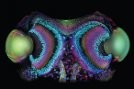
Figure 2 - First-place winner: Eyes of Daddy Longlegs (Harvestman).
First place went to Dr. Igor Siwanowicz, amusingly self-proclaimed “Ambassador of Bugs” of the Max Planck Institute for Neurobiology (Munich, Germany). He captured a confocal microscope image that showcases the bug-eyed splendor of a Daddy Longlegs, also known as a Harvestman or Phalangium opilio. The photo reveals not only the eyes’ lenses (two large ovals), but also the retinas and optic nerves (trailing down at center back) (Figure 2). The reward for first place was $5000 worth of Olympus equipment.
Nearly 2000 entries had participated in the 2010 competition, providing images and movies that captured a wide variety of samples, many of which were showcased in a vibrant video displayed during the award celebration.
To continue reading about the other winners and honorable mentions, please visit 2010 Olympus BioScapes Digital Imaging Competition: Winners and Honorable Mentions.
Conclusion
Although we have been looking at cells since the 17th century, much remains to be seen and discovered. There are approximately 60–90 trillion cells in the adult human body—enough cells to line up together to circle the earth almost five times. The ASCB Annual Meeting showcased a tremendous amount of new research and emerging technologies that will enable us to look further into the extraordinary complexity of the cell.
For more information about the ASCB Annual Meeting and to read full abstracts, please visit http://www.ascb.org/meetings/.
Next year, the ASCB 51st Annual Meeting is scheduled to be held December 3–7, 2011, in Denver, CO. For more information about the American Society for Cell Biology, please visit http://www.ascb.org.
Ms. Hunt is Content Editor for American Laboratory/Labcompare; e-mail: [email protected].