Surfaces, the two-dimensional outer boundary between the three-dimensional object and its environment, have many attributes and are perceived as the complex interplay of three factors: surrounding illumination, surface optical properties such as reflection, and surface geometry or topography.1 Interest in the characteristics and nature of the surface geometry of photographs is growing in scientific and museum circles. Surface metrology, the measurement of areal features of roughness and waviness from surface textures, is well suited for the study of cultural heritage, historic, and artistic works as a means to characterize their surfaces. Advances in electronics, optics, and faster computing in the past 10 years have revolutionized the field of optical and surface metrology. Earlier surface metrology contact methodologies, which irreversibly perturb surfaces, were not safe for the study of priceless art and cultural heritage objects. New faster optical surface metrology tools such as confocal scanning disk microscopy use light to probe surfaces rapidly and without risk, and yield a wealth of areal data unachievable with earlier contact methods. Such instrumentation enables nonperturbing (noncontact, nondestructive, and noninvasive) examination of delicate and sensitive surfaces of the wide range of historic and artistic works encompassing the cultural heritage accumulated by humankind. These new surface metrology instruments open up museum depositories for a new type of collection- based studies.2-7
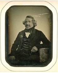
Figure 1 - Portrait of Louis-Jacques-Mandé Daguerre taken in 1844 by French daguerreotypist Jean-Baptiste Sabatier-Blot. George Eastman House collection, catalog number 1976:0168:0043, quarter plate (69 × 91 mm2).
Since their inception, photographs have been generated using a wide range of materials such as silver, mercury, and gold for daguerreotypes; egg proteins for albumins; cellulose nitrate for collodions; acacia gum for gum prints; and gelatin for silver gelatin prints. The daguerreotype, the first commercially viable photographic system, was introduced to the world in Paris in 1839 by Louis-Jacques-Mandé Daguerre (1787-1851). Figure 1 shows a daguerreotype portrait of Monsieur Daguerre taken in 1844 by fellow Frenchman Jean-Baptiste Sabatier-Blot (1801-1881). Daguerreotypy deeply impressed the world and it rapidly spread to Europe, America, and other destinations around the globe, and millions were made during its heyday from 1839 to 1860. In addition to the daguerreotype's overwhelming popular appeal and because of its primacy in the history of photography, daguerreotypes were first to document people and places: They provided scientific, social, anthropological, and archaeological images to the public and scholars of the era. Today, as silver-based photography has given way to silicon and digital imaging, the daguerreotype is increasingly treasured for its fine art, artefactual, and documentary potential.
This paper presents surface characterization studies using confocal scanning disk microscopy of three daguerreotypes—two from the George Eastman House International Museum of Photography and Film collections, and one from the Public Library of Cincinnati and Hamilton County (OH).
The confocal principle developed by Marvin Minsky8 in 1955 is a microscopic imaging system that uses a spatial pinhole filter to eliminate out-of-focus light in specimens that are thicker than the focal plane. This concept was not seriously considered until 20 years later, when it was realized that the depth or z resolution is greater than that of optical microscopy at the expense of a smaller field of view. This optical sectioning or depth-discrimination property is its strength—its ability to capture the depth of a sample and image in the z direction at various heights, to yield a full 3-D image from a stack of partial images.9,10
Experimental methods and materials
In this research, a µsurf optical 3-D measurement system (NanoFocus AG, Oberhausen, Germany) based on the confocal scanning disk principle was used with 10× and 20× objectives. Data acquired from the system only provide information on the geometry and physical structure of a surface and not its chemical composition. The confocal system provides 3-D surface data arrays of 512 × 512 points for all objectives, represented graphically by contour and isometric maps. A 10× objective with a numerical aperture (NA) of 0.30 and a working distance (WD) of 11 mm provided a 1.55 × 1.55 mm2 measurement field with a vertical resolution of 50 nm, and a 20× objective with NA 0.60, WD 0.9 mm, 800 × 800 µm2 field, and vertical resolution of 4 nm. Version 6.1 of µsoft software was used for operation of the confocal topometer, generating contour and isometric images and profile views. µsoft Analysis Standard software version 4.1 was used for data analysis and calculation of surface texture height parameters: arithmetic mean deviations, Sa, and root-mean-square (RMS) deviations, Sq, of the surface.
Three daguerreotypes were examined in this study. Two daguerreotypes, from the George Eastman House Department of Photography collection, are portraits made by the well-known Boston daguerreotype studio of Albert Sands Southworth (1811-1894) and Josiah Johnson Hawes (1808-1901), and are whole plates measuring 165 × 216 mm2: 1) unidentified man, circa 1850, catalog number 1974:0193:0621; and 2) Boston's notable physician and abolitionist Henry I. Bowditch, seated, circa 1850, catalog number 1974:0193:0043. The third daguerreotype examined is plate number 3 of the eight whole plates that make up the Cincinnati waterfront panorama photographed by Charles Fontayne and William S. Porter in 1848, now part of the collection of the Public Library of Cincinnati and Hamilton County.
Results and discussion
The daguerreotype imaging system is unique in that only one photograph of high resolution is produced. As with later photographic and current imaging systems, multiple prints are not feasible. Another unique daguerreotype feature is that the image generated rests on the surface of a highly polished silver plate: The image is the surface and vice versa.11 The Cincinnati waterfront panorama clearly shows this phenomenon as well as the high resolution of this first photographic system.
Cincinnati waterfront panorama, plate 3
The Fontayne and Porter Cincinnati waterfront panorama was photographed from about 2 km away in the town of Newport, KY, on the southern shore of the Ohio River. Three years after it was taken, it traveled across the Atlantic to be exhibited at The Great Exhibition of 1851, a showcase of British industrial, technical, economic, and military prowess, held at the Crystal Palace in Hyde Park, London. The panorama of this frontier city was greatly admired, and both photographers won awards for their technical achievement with this new medium.

Figure 2 - Plate 3 of the Cincinnati Waterfront
eight-plate panorama daguerreotype, taken by Fontayne and Porter in 1848. Public Library of Cincinnati and Hamilton County collection, whole plate (165 × 216 mm2). Arrow indicates area studied.
Each of the eight whole plates measures approximately 165 × 216 mm2. Plate 3, shown in Figure 2, presents a portion of the river waterfront lined with numerous paddle steamboats, buildings and houses, and tree-covered hills in the background. It also shows the current condition of the plate, a brown tarnish along all borders delineating the position of the display paper mat. Figure 3 shows the results of examining the name on the protective cover of the paddle wheel on one of the steamboats with optical and confocal microscopy, indicated by the blue arrow in Figure 2. The top image on Figure 3 shows the “WA” on the protective cover of the paddle wheel. This demonstrates the high resolving power of the daguerreian medium when considering that the image was taken from about 2 km away across the Ohio River. The two lower images are of the “W” taken with the confocal spinning disk microscope. The lower left image is a false color contour plot showing the relief of the “W” and the lower right image is a 3-D isometric plot in which red, yellow, and white represent higher areas and peaks, and green and blue are lower regions. This is in accordance with the physical nature of the daguerreotype image and surface: 1) The white areas are composed of light scattering image particles with a size range from 200 to 800 nm; these appear as the yellow peaks clearly observed in the 3-D isometric plot. 2) The dark areas correspond to the highly polished plate areas with few to no image particles that are beneath the white image particle area and show off the green “W” in the isometric plot.

Figure 3 - Detail from the paddle cover of the steamboat in Figure 2 demonstrating the image to be a surface phenomenon: image = surface.
Unidentified man, whole plate
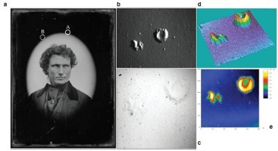
Figure 4 - a) Daguerreotype portrait of unidentified man by studio of Southworth and Hawes, about 1848–50 (×20 objective power, NA: 0.60) (area A shows metallic blisters; area B shows a large exfoliated area); b) blisters obliquely illuminated at 100× magnification; c) blisters in reflected light, brightfield; d) isometric view in gray scale of blisters from confocal 3-D data; e) contour or topographic view in gray scale of blisters from confocal 3-D data.
The portrait of an unidentified man was examined with stereo-, optical, and confocal microscopy. From a distant perspective, the plate appeared to be in good condition. Close examination revealed small surface features such as metallic protrusions (blisters) (area A in Figure 4a) and exfoliated areas (loss of surface metal) (area B in Figure 4a) throughout the top portion of the plate.12
Blisters
The set of blisters in area A (Figure 4a),
and detailed in Figure 4b–e, are located
above the man’s head and were captured
with various microscopic modalities. Figure
4b is a stereoscopic image of the blisters
obliquely illuminated at 100× magnification
that qualitatively shows the 3-D nature of the blisters. Figure 4c is a
reflected light, brightfield image of the
blisters that does not accentuate the 3-D
nature of these blisters.

Figure 5 a) Contour image with b) profile running through small blister on the right and large, crater-like
blister (heights and widths of blister are listed in μm).
Figure 4d, a 3-D isometric plot in gray scale,
and Figure 4e, a contour plot in gray scale,
clearly illustrate the 3-D nature of these blisters.
From the confocal data, quantitative
physical information can be extracted to
reveal such features as the height of blister
peaks and the width or diameter of blisters.
Figure 5a shows a contour view with a black
line profiling the large blister on the top right.
A profile (Figure 5b) shows the heights of the
single peak (0.622 mm) of the small blister on
the left, the doublet (1.520 and 1.226 mm) of
the large blister on the right, and the widths
of the two peaks: The small peak is 35.9 mm,
and the large doublet is 271 mm. The z-axis
features are considerably smaller than the lateral
features. For example, the small peak on
the right side of the profile is 0.622 mm high
versus its width of 35.9 mm.